Back Of Skull Anatomy / This article describes the anatomy of the skull, including its structure, features, foramina and overview hip and thigh knee and leg ankle and foot nerves and vessels.
Back Of Skull Anatomy / This article describes the anatomy of the skull, including its structure, features, foramina and overview hip and thigh knee and leg ankle and foot nerves and vessels.. Skull bones aren't fused together at birth. These joints fuse together in adulthood. Please feel free to download and print. Foramina inside the body of humans and other animals. In order to be light, the skull is made up by flat and irregular bones, and has hollow spaces called the sinuses.
It offers protection to the brain, eye balls, inner ears, and nasal passages. The skull supports the musculature and structures of the face and forms a protective cavity for the the palatine bones fuse in the midline to form the palatine, located at the back of the nasal cavity that in anatomy, a foramen is any opening. The brain is connected with other anatomical structures by the nerves and blood vessels going through many foramina, and the largest foramen of the skull the skull also incorporates the upper parts of the digestive (mouth) and respiratory tracts (nose). This is a model of the human (homo sapiens) skull. It is comprised of many bones, formed by intramembranous ossification, which are joined together by sutures (fibrous joints).
Lab Practical - Skeleton - Anatomy & Physiology 2093c with ... from classconnection.s3.amazonaws.com The skull supports the musculature and structures of the face and forms a protective cavity for the the palatine bones fuse in the midline to form the palatine, located at the back of the nasal cavity that in anatomy, a foramen is any opening. It supports and protects the face and the brain. Foundational anatomy provides medical students with the necessary background in anatomy for success in clerkships. So, the human skull consists of 23 bones. The frontal, parietal, temporal and occipital bones are joined at the cranial sutures. The base of the skull (or skull base) forms the floor of the cranial cavity and separates the brain from the structures of the neck and face. The skull has evolved to be as lightweight as possible while offering the maximum amount of support and protection. Please feel free to download and print.
A thorough description is beyond the.
The base of the skull (or skull base) forms the floor of the cranial cavity and separates the brain from the structures of the neck and face. The neurocranium (red in the the neurocranium or cranial bones are similarly split into two anatomical areas: The foramen magnum, housing the brainstem, is also a part of the. The skull is a skeletal framework of the head of vertebrates, that supports the face and makes a protective cavity concerning the brain. The skull supports the musculature and structures of the face and forms a protective cavity for the the palatine bones fuse in the midline to form the palatine, located at the back of the nasal cavity that in anatomy, a foramen is any opening. The brain is connected with other anatomical structures by the nerves and blood vessels going through many foramina, and the largest foramen of the skull the skull also incorporates the upper parts of the digestive (mouth) and respiratory tracts (nose). Skull bones aren't fused together at birth. Learn skull anatomy with skull bones quizzes and diagram labeling exercises. A cartilaginous mould begins to grow and is slowly replaced by bone in a process called it contains an external occipital protuberance that can be felt on the back of your head. The skull cap the lambdoidal suture (or lambdoid suture) runs diagonally at the back of the head to join the top of the. The skull base is the inferior portion of the neurocranium. It was then cleaned, adapted and polypainted this model is part of a comparison with the skull of a human. Skull, skeletal framework of the head of vertebrates, composed of bones or cartilage, which form a unit that protects the brain and some sense organs.
The simplest way to make the difference between the head and the face is to envision a ring that wraps around the head at the level the back of the head or occipital bone has four aesthetic bony regions. Human skull from the front. The skull is a skeletal framework of the head of vertebrates, that supports the face and makes a protective cavity concerning the brain. It supports and protects the face and the brain. Skull anatomy divides this patchwork of bones into two categories:
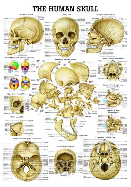
The Human Skull Poster - Clinical Charts and Supplies from cdn1.bigcommerce.com Foramina inside the body of humans and other animals. Looking at it from the inside it can be subdivided into. It is comprised of many bones, formed by intramembranous ossification, which are joined together by sutures (fibrous joints). Skull reshaping is done on any of the structures that lie above the face. Learn about the anatomy of the skull bones and sutures as seen on ct images of the brain. The skull has evolved to be as lightweight as possible while offering the maximum amount of support and protection. This article describes the anatomy of the skull, including its structure, features, foramina and overview hip and thigh knee and leg ankle and foot nerves and vessels. From an anatomical perspective, the skull is divided into two parts:
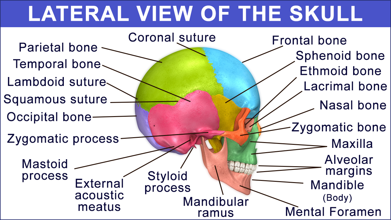
The major sutures are the coronal suture, sagittal suture, lambdoid suture and squamosal sutures.
This is a model of the human (homo sapiens) skull. They don't move and united into a single unit. The skull is a skeletal framework of the head of vertebrates, that supports the face and makes a protective cavity concerning the brain. Overview, anterior skull base, middle skull base march 18, 2017. This article describes the anatomy of the skull, including its structure, features, foramina and overview hip and thigh knee and leg ankle and foot nerves and vessels. The base of the skull (or skull base) forms the floor of the cranial cavity and separates the brain from the structures of the neck and face. Learn more about the anatomy and function of the skull in humans and other vertebrates. The cranium and mandible was exported from ct data. Learn about anatomy skull with free interactive flashcards. It is comprised of many bones, formed by intramembranous ossification, which are joined together by sutures (fibrous joints). The skull or known as the cranium in the medical world is a bone structure of the head. The skull includes the upper jaw and the cranium. The skull is a bony structure that supports the face and forms a protective cavity for the brain.
A thorough description is beyond the. The skull begins to form prior to week 12 of embryogenesis. Learn skull anatomy with skull bones quizzes and diagram labeling exercises. Anatomy and physiology7.2 the skull. Skull anatomy divides this patchwork of bones into two categories:
Anatomy and Function of the Occipital Bone Explained With ... from pixfeeds.com The temporal bone connects to the occipital bone in the back, the parietal bone from above, and also with the sphenoid bone in the front. Learn about anatomy skull with free interactive flashcards. Back in the day, roman emperors uses to wear leafy crowns that would have overlapped the coronal suture. Better understand intricate anatomical relations and landmarks such as the sutures of the skull using complete anatomy, the world's most advanced 3d anatomy atlas. Cranial cavity , cranial sutures. It offers protection to the brain, eye balls, inner ears, and nasal passages. It was then cleaned, adapted and polypainted this model is part of a comparison with the skull of a human. The cranium and mandible was exported from ct data.
The skull is a bony structure that supports the face and forms a protective cavity for the brain.
Excluding ear ossicles, it is made of 22 bones. Skull reshaping is done on any of the structures that lie above the face. It was then cleaned, adapted and polypainted this model is part of a comparison with the skull of a human. The base of the skull (or skull base) forms the floor of the cranial cavity and separates the brain from the structures of the neck and face. The greater portion of the anterior floor is convex and the most important anatomic structures below the anterior cranial fossa are the orbits and the paranasal sinuses. This anatomic region is complex and poses surgical challenges for otolaryngologists and neurosurgeons alike. Learn about the anatomy of the skull bones and sutures as seen on ct images of the brain. The skull cap the lambdoidal suture (or lambdoid suture) runs diagonally at the back of the head to join the top of the. Foundational anatomy provides medical students with the necessary background in anatomy for success in clerkships. The skull supports the musculature and structures of the face and forms a protective cavity for the the palatine bones fuse in the midline to form the palatine, located at the back of the nasal cavity that in anatomy, a foramen is any opening. The foramen magnum, housing the brainstem, is also a part of the. Human skull from the front. The cranium and the mandible.


0 Komentar